Please note that the Knowledge Hub is purely for reference and that you do need to enquire with either your medical practitioner or a certified doctor for proper diagnosis.

Fractures of the bones of the ankle (tibia, fibula and talus). This usually occurs acutely with a traumatic event (e.g., a fall).
Ankle pain, swelling, deformity and an inability to weight bear. Xrays, CT scan, MRI scan.
Loss of cartilage of the ankle joint (cartilage of the tibia and/or talus). This results in ankle pain, swelling, clicking, deformity and instability. The cartilage loss is due to an underlying inflammatory condition such gout, rheumatoid arthritis, psoriasis and infection.
Ankle pain, swelling, tenderness and instability. Painful weight-bearing. Xray’s, CT scan or MRI scan.
Loss of cartilage of the ankle joint (cartilage of the tibia and/or talus). This results in ankle pain, swelling, clicking, deformity and instability. The cartilage loss is usually from cartilage ‘wear & tear’.
Ankle pain, swelling, tenderness and instability. Painful weight-bearing. Xrays, CT scan or MRI scan.
Injuries to the tendons of the ankle. This can occur acutely with a traumatic event of over time due to a repetitive activity. The most common tendon injuries around the ankle are the Achilles tendon, Peroneus tendons (Peroneus Brevis & Peroneus Longus) and the Posterior Tibialis tendon.
Pain, swelling and an inability to weight bear. Xray’s, Ultrasound and MRI scan

Also known as ulnar nerve neuropathy. This is a compression of the ulnar nerve around the inner aspect of the elbow (most commonly at the elbow medial epicondyle – the bony prominence on the inner aspect of the elbow). This results in pain, numbness and ‘pins & needles’ along the ulnar nerve distribution in the forearm and hand (inner aspect of forearm and pinkie and ring fingers).
Classic neurological symptoms along the ulnar nerve in the forearm and hand (inner aspect of the forearm, pinkie and ring fingers). Symptoms include pain, numbness and ‘pins & needles’. Referral to a neurologist for Ulnar nerve conduction studies and EMG (electromyography).
The distal biceps tendon is pulled off its attachment on the radial tuberosity of the forearm. The patient will have weakness with elbow flexion and forearm supination. This is an acute injury and is associated with elbow swelling and bruising.
Elbow bruising and elbow swelling. Weak elbow flexion and elbow supination. Ultrasound and MRI scan
Fractures around the elbow are very common, especially in children.
Fractures are diagnosed with Xray’s and/or CT scan.
Also known as medial epicondylitis. This is an overuse injury of the flexor tendons of the wrist & fingers resulting in pain over the medial epicondyle of the humerus (the bony prominence on the inner aspect of the elbow).
Pain and tenderness over the medial epicondyle (bony prominence on the inner aspect of the elbow). Ultrasound
Also known as lateral epicondylitis. This is an overuse injury of the extensor tendons of the wrist & fingers resulting in pain over the lateral epicondyle of the humerus (the bony prominence on the outer aspect of the elbow).
Pain and tenderness over the lateral epicondyle (bony prominence on the outer aspect of the elbow). Ultrasound

Acute and chronic inflammation of the Achilles tendon. This may cause pain, swelling and thickening of the Achilles tendon. Pain may be worsened when walking on the toes of the affected foot.
Achilles tendon pain, tenderness and swelling. Ultrasound
A bunion is a bony bump on the outside of the big toe. This occurs when some of the bones of the forefoot move out of place. The big toe deviates to the lesser toes and the big toe joint forms a bony bump on the outside. The skin over this bump may be stretched and inflamed causing pain/tenderness.
Clinical features. Xrays
Usually affects the big toe. The sides of the toe nail grows into its soft tissue edge. This can cause significant inflammation of the soft tissue surrounding the nail (redness, warmth and tenderness).
Inflammation of the toe in the presence of an ingrown toenail.
A thickening of the digital nerve (neuroma) in the forefoot, most often in the 3rd This may cause a sharp pain in the forefoot with weight bearing. The pain may be associated with toe numbness and/or pins & needles most often in the 3rd forefoot web space.
Sharp pain in the forefoot webspace. The pain is worsened with compression of the forefoot. Ultrasound and MRI scan.
The plantar fascia is a thick fibrous band in the sole of the foot that runs from the toes to the heel. Inflammation of the plantar fascia is one of the most common causes of heel pain.
Heel pain where the plantar fascia attaches to the heel. Pain is experienced with weightbearing, especially in the morning (first few steps). Xrays often show a bony heel spur where the plantar fascia is attached.

Bursae are small membranes that lie at muscle-bone interfaces, preventing friction. Bursae can become inflamed and cause significant pain (this is called bursitis). Hip greater trochanteric bursitis is very common.
Pain over the hip greater trochanter, Ultrasound or MRI
A condition where the femoral head pops out of the socket.
Sudden onset acute groin pain and an inability to walk/stand. Xrays, CT scan or MRI
Hip development problem at birth. The ball & socket hip joint has not developed correctly resulting in hip pain.
Groin pain, Xrays or MRI
A fracture of the femoral head and/or femoral neck and/or femoral socket.
Sudden acute pain and inability to walk/stand. X-rays, CT scan or MRI scan
Also known as Femoro-acetabular impingement. A condition where the femoral head makes contact with the acetabulum (socket). The head and socket impinge. This can cause significant hip pain with hip movement.
X-rays or MRI arthrogram
Loss of cartilage of the hip joint (cartilage of the femoral head and/or socket). This results in groin pain with hip movements. The cartilage loss is due to an inflammatory condition such as gout, rheumatoid arthritis, psoriasis and infection. Medical management of the underlying inflammatory condition.
Groin pain, Blood tests (inflammatory markers, X-rays or MRI)
The hip labrum is a thick cartilage structure circumferentially on the rim of the hip socket. The main function of the labrum is to provide hip stability, protect the cartilage of the socket and femoral head, increase the socket surface area and provide a ‘suction’ effect on the femoral head. When the labrum tears or detaches from the socket rim this is called a labral injury.
Groin pain/clicking, MRI Arthrogram
Ligament injury of the hip. Injuries can be acute (sport injuries or accidents) or chronic (degenerative tears). Most common hip ligament injuries are the Ligamentum Teres and the Ilio-femoral ligaments.
Groin pain, hip muscle weakness. MRI scan
Muscle injury of the hip. Injuries can be acute (sport injuries or accidents) or chronic (degenerative tears). Most common hip muscle injuries are the abductor muscles (Gluteus Medius and Gluteus Minimus) and hip flexor muscles (iliopsoas muscle).
Groin pain, hip muscle weakness. MRI scan
Loss of cartilage of the hip joint (cartilage of the femoral head and/or socket). This results in groin pain with hip movements. The cartilage loss is due to ‘wear & tear’.
Groin pain, Xray or MRI

Bursae are small membranes that lie at muscle-bone interfaces, preventing friction. Bursae can become inflamed and cause significant pain (this is called bursitis). The most common knee bursitis is prepatellar bursitis (housemaids knee), suprapatellar bursitis, infrapatellar bursitis and pes anserine bursitis.
Knee pain and swelling with direct palpation. Ultrasound, MRI
Fractures around the knee (distal femur, proximal tibia and knee cap). This occurs as a result of a traumatic event (e.g., fall).
Significant knee pain and swelling and an inability to walk. X-rays, CT scan, MRI scan
Loss of Cartilage from the articulating knee surfaces (patella, femur and/or tibia). This can result in knee pain, swelling, deformity, crepitations and locking. This is usually due to an underlying inflammatory condition such as gout, rheumatoid arthritis, psoriasis and infection.
Knee pain, swelling, deformity, locking and instability. X-rays.
Injury to the knee ligaments. This usually occurs as an acute event after an accident or sport injury. The most common knee ligament injuries are ACL (anterior cruciate ligament), PCL (posterior cruciate ligament), MCL (medial collateral ligament) and LCL (lateral collateral ligament) injuries. Knee ligament injuries result in knee pain, swelling and instability.
Knee pain, swelling and instability following a traumatic event. Xrays, Ultrasound, MRI
Muscle injuries adjacent to the knee. Most common muscle injuries around the knee are quadriceps, hamstring and Iliotibial band injuries (ITB). This is usually associated with sport or trauma.
Knee pain and weakness usually associated with an identifiable event. Ultrasound, MRI
Loss of Cartilage from the articulating knee surfaces (patella, femur and/or tibia). This can result in knee pain, swelling, deformity, crepitations and locking. This is usually due to ‘wear & tear’ of the cartilage.
Knee pain, swelling, deformity, locking and instability. X-rays.
Injury to tendons in the knee. This can be as a result of a sudden acute traumatic event or can be due to a repetitive sprain injury. The most common knee tendon injuries are patella tendon injuries, MPFL (medial patella-femoral ligament) and quadriceps tendon injuries.
Knee pain, swelling and tenderness. Pain with direct tendon palpation. Ultrasound, MRI
Destruction of cartilage under the kneecap. This may be due to an acute traumatic event or as a result of a chronic patella disorder (see above). The cartilage destruction is often described as having a ‘crab meat’ appearance.
Pain, swelling and crepitations under the knee cap, this may be associated with swelling. Xrays, MRI scan
Abnormal development of the patella. The most common patella disorders are poor patella tracking, abnormal patella shape (dysplasia), a patella that lies too far above the knee joint (patella alta) and a patella that lies too far below the knee joint (patella baja).
Pain and clicking under the knee cap and knee swelling. X-rays, CT scan and MRI scan

Fractures of the pelvic region. This includes fractures of the 3 pelvic bones (ilium, ischium and pubis). The acetabulum is also known as the hip socket (pelvic socket). Acetabulum fractures occur with significant traumatic forces. These fractures require urgent attention as neglect can result in hip osteoarthritis.
Pelvic Xray’s, CT scan
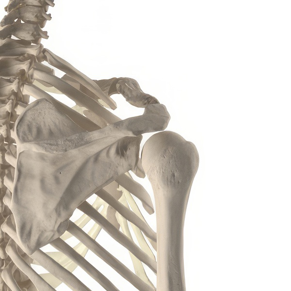
Dislocation of the acromio-clavicular joint. This is due to a traumatic event (sport/accident). The relationship between the acromion and the clavicle is disturbed.
An identifiable traumatic event. Acute onset pain in the AC joint. Xrays.
Also known as Rotator cuff impingement. The rotator cuff is the name given to the group of muscles of the shoulder. The rotator cuff may impinge on the acromion (the bone just above the shoulder). This will result in pain with shoulder movement and may cause rotator cuff tears.
Shoulder pain with range of motion (especially overhead activity). Xray’s, Ultrasound and MRI scan.
Loss of cartilage from the articular surface of the shoulder (humeral head and glenoid). This loss of cartilage may be due to an underlying inflammatory condition like gout, rheumatoid arthritis, psoriasis and infection.
Progressive shoulder pain, swelling, stiffness and crepitations. Xrays & MRI scan.
An unstable shoulder that dislocates (pops out of its socket). Patients avoid certain shoulder positions for fear of dislocation.
Patient reports serial shoulder dislocations. Xrays, CT scan, MRI scan.
Loss of cartilage from the articular surface of the shoulder (humeral head and glenoid). This loss of cartilage is usually due to ‘wear & tear’.
Progressive shoulder pain, stiffness and crepitations. Xrays & MRI scan

Carpal tunnel syndrome is compression of the median nerve in the wrist (the wrist is also known as carpus). This results in pain, numbness and a tingling sensation in the thumb, index, middle and ring fingers). Patients may be kept awake at night with the above-mentioned symptoms. This may be associated with, hypothyroidism and pregnancy.
Referral to a neurologist for Ulnar nerve conduction studies and EMG (electromyography).
A painful condition affecting the thumb. The tendons on the thumb side of the wrist are inflamed, this causes pain with thumb activities such as opening a bottle/tap and wringing a cloth.
Pain and swelling along the tendons of the thumb at the wrist. Passive flexion of the thumb is painful. Ultrasound
A cyst that originates from a joint or tendon. The cyst is filled with a gel-like fluid. A ganglion is a non-cancerous. Ganglions are most commonly found around the wrist.
Clinical appearance of the ganglion (soft fluid filled sac). Ultrasound
Ulnar nerve compression at the pinkie side of the wrist. This may cause wrist pain and numbness or tingling in the pinkie and ring fingers.
Numbness and/or tingling of the pinkie and ring finger. Referral to a neurologist for Ulnar nerve conduction studies and EMG (electromyography).
The flexor tendon of a finger is stuck when the finger is flexed (bent into the palm). The finger is then straightened with a snap (like releasing a trigger).
The diagnosis is made clinically but can be confirmed with an Ultrasound.


The information provided on this website is for educational purposes only and is not intended as a substitute for professional medical advice, diagnosis, or treatment. Always seek the advice of your physician or other qualified health provider with any questions you may have regarding a medical condition. Never disregard professional medical advice or delay in seeking it because of something you have read on this website. If you think you may have a medical emergency, call your doctor or emergency services immediately. The content on this website is based on the author’s opinions and is not intended to be a comprehensive treatment of the subject. While we strive to provide accurate and up-to-date information, we make no guarantees regarding the completeness, reliability, or accuracy of the information on this site. Links to other websites are for informational purposes only and do not constitute an endorsement of those sites. We are not responsible for the content of any external websites. By using this website, you acknowledge that you have read and understood this disclaimer and agree to its terms.